Thursday, November 5, 2015

Periapical lesions, when they grow big and stubborn


Inflammatory lesions of dental pulp infection origin are the most common periradicular lesions of the jaws. Endodontic lesions may be classified into periapical granulomas, chronic apical abscesses and periapical cysts. These three endodontic lesions are usually unilocular, periapical radiolucencies of varying in sizes and marginal definition.

The differential diagnosis for large endodontic lesions will include odontogenic keratocysts, ameloblastomas, lateral periodontal cysts, nasopalatine duct cysts and periapical fibrous scars.

Root canal treatment (RCT) or retreatment is the first line of treatment for large endodontic lesions. If these lesions do not resolve after RCT, then apical surgery would be indicated. Other indications of apical surgery include failed retreatment cases due to calcified canals, procedural errors, eg. separated instruments or any obstructions that prevent thorough biomechanical preparation of the root canal system, apical transportation, foreign body or excessive root filling material in the periapical lesion and/or the presence of post restorations. 


Periapical inflammation is a direct effect of bacteria infection of the root canal system (Kakeshashi et al, 1965; Moller et al, 1981). The inflammation is a result of interaction between bacteria in untreated infected root canal systems and the host’s defense or immune system. Inadequate infection control of the primary infection is the main reason for the failed RCT, leading to persisting periapical inflammation.

Studies have shown that non-surgical root canal treatment failed because bacteria can survive and maintain an infectious disease in periapical lesions, although the size of the lesions were not mentioned (Tronstad et al, 1987; Tronstad et al, 1990; Sunde at al, 2002). The microbiota of persistent apical periodontitis lesions is composed of various types of microorganisms with biofilm-forming capacity. The majority of the bacterial strains of extraradicular infection were facultative anaerobes (51.6%) and obligate anaerobic bacteria (38.7%). Bacteria were also found in abscesses and cysts of extracted teeth with lesions attached to the apices (Ricucci et al, 2006). Actinomycotic infections were found to be more common in periapical infections than previously believed and apical surgery including curettage of the infected area with resection of the involved roots is the treatment of choice.  

From a histological point of view, large periapical lesions may either be granulomatous or cystic. Both periapical granulomata and cysts have the same clinical and radiographic appearance. It is difficult to differentiate between the two lesions based on radiographic density of the periapical lesion. A very well defined lining on the radiograph only indicates a long-standing lesion that is slowly increasing in size, whereas a diffuse border indicates a rapidly expanding lesion. The size of the radiolucency is also irrelevant to the histological state of the tissue as both small and large lesions can be granulomata, abscesses or cysts.

The progression of apical periodontitis to periapical cyst may be seen as an end stage of an ongoing host defense response to root canal infection. A periapical cyst is believed to be a direct sequel of a periapical granuloma. The transition from granuloma to cyst is based on gradual increasing proportions of epithelium and development of a defined lumen. Although there are several theories proposed, little is known of the pathogenesis of cysts. The prevalence of periapical cysts among apical periodontitis lesions ranges from 8.7% to 37.7%, using different criteria in histological studies. Granulation tissue with epithelial proliferation without cavitation is an insufficient diagnosis of a cyst, and is classified as granuloma. Based on strict histological criteria used by Nair et al (1996), cystic formation are often in the minority and may constitute 15% of lesions studied, 9% true cysts and 6% periapical pocket cysts. As the pocket cyst is directly connected to the root canal system, healing is likely to occur after removal of intracanal bacteria during RCT. A true cyst is self-sustaining because the lumen of a true cyst is completely enclosed by the epithelium and there is no communication with the root canal system. Since the cyst is independent on presence of absence of irritant in the root canal, it is not likely to resolve by RCT and thus surgical intervention is necessary.


Retreatment was carried out prior to surgery.  Biomechanical instrumentation reduces microorganisms and their toxins in the root canal system. Together with a good quality root canal filling and a retrofill with a tight seal, the egress of microorganisms and their toxins from the canals into the periapical tissues will be kept to a minimum. This promotes healing and ultimately, reduce the incidence of healed lesions relapsing.

To ensure a good outcome of an apical surgery, the main treatment concepts are outlined below:

1.     Magnification and illumination using the dental operating microscope
The outcome of endodontic surgery was compared between the use of dental operating microscope and loupes or no magnification, in a meta-analysis of 101 articles from the endodontic literature (Setzer et al, 2012). The probability for success for endodontic microsurgery proved to be significantly greater than the probability for success for conventional root end surgery, providing best available evidence on the influence of high-power magnification rendered by the dental operating microscope or the endoscope.

2.     Achieving profound hemostasis
Hemostasis is essential for better visualization during apical surgery to create a dry surgical site to facilitate the insertion of root end fillings and reduce post-operative bleeding. Vasoconstrictors in local anesthetics reduce the blood flow at the surgical site. The use of 1:50000, or at least 1:80000 epinephrine (as in Articaine) is recommended. Cotton pellet with epinephrine and ferric sulphate can effectively control bleeding.

3.     Always resect the root and retrofill
Resection of 3mm of the root tip reduces more than 90% of the apical ramifications and lateral canals, without compromising on the strength and stability of the tooth. Root end fillings placed after root resection shows greater healing success compared to those with only root resection. The root end cavity preparation should be at least 3mm into the root dentin. The ultrasonic angled retrotips provide easier access to the root end.

4.     Choice of retrofilling material
This ideal root end filling material should be biocompatible, regenerates periapical tissues and has excellent sealing ability. Amalgam as the traditional retrofill material is unacceptable nowadays because its sealing ability is questionable, it corrodes and tattoos the gingiva or mucosa. Mineral trioxide aggregate (MTA) and zinc oxide eugenol containing materials, such as IRM, are the more commonly used materials. MTA is extensively researched in endodontics and it is shown to be biocompatible and bioactive when set and produces a high pH before setting, which results in regeneration of periapical tissues including cementum and periodontal ligaments. However, this material requires mixing and not easy to manipulate. The endodontic pre-mixed bioceramics (Totalfill BC RRM-Putty) contains calcium silicates, zirconium oxide, calcium phosphate, tantalum oxide and fillers; has excellent mechanical and biological properties; and easy to handle.


The resected tooth was checked for clinical signs of persisting infection in every six monthly review session. Radiographic signs of density change within the lesion, trabecular reformation and lamina dura formation are evidence of periapical healing. Scar tissue is a reparative response, producing fibrous connective tissue instead of bone. Scars occurred more frequently when both the buccal and lingual cortical plates are lost. Formation of the scar tissue should not be interpreted radiographically as an endondontic treatment failure or misdiagnosed as an infected lesion. A long-term review up to four years is essential to confirm successful outcome of the apical surgery of these teeth.


Friday, October 2, 2015

Hola Espana!

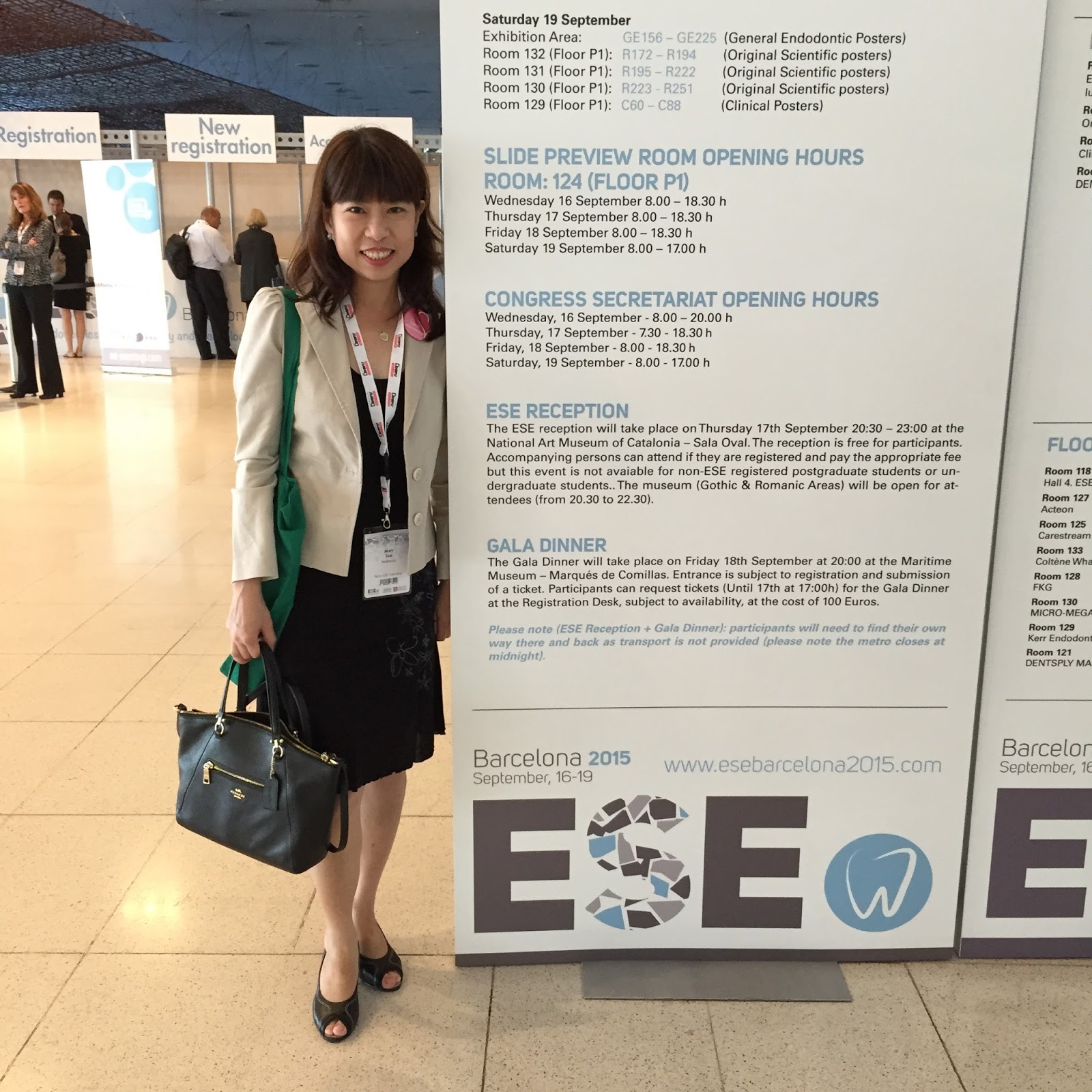
This is my first ESE (European Society of Endodontology) Biennial Congress at Barcelona. It was attended by more than 3000 endodontists/dentists/postgraduate students from all round the world. Dental conferences like this one is perfect to catch up with old friends. Old ties renewed and some familiar faces reappeared. Barcelona is really a beautiful city with great beaches, delicious seafood tapas and friendly Spanish speaking people, and that made my 14-hour long flight from Kuala Lumpur worth enduring...


Tuesday, April 14, 2015

Peri-radicular healing


As a dental healthcare provider, I get self-satisfaction not only when my patients are happy with me, but when I see good results from my own work, especially when it comes to challenging root canal treatment. The radiograph showed periapical healing after one year, a favorable outcome of the root canal treatment for tooth 36 with chronic apical abscess. The evidence of bone filling the peri-radicular lesion never failed to put a smile on my face :)


The second case shows tooth 35 after apexification with MTA and periapical healing after nine months.

Sunday, June 15, 2014

Digital radiography: Before & After


C-shaped canal in tooth 36 with large peri-radicular lesion, buccal swelling and deep lingual pocket. Three months review pa showed resolution of the lesion.


Retreatment on tooth 46 with underprepared canals and short root fillings.


Root canal treatment on tooth 26.

These images were taken using the Carestream RVG6100. Digital radiography provides much more clarity, with instant access to the images.


Wednesday, April 16, 2014

Happy Birthday Pa!


My dad will be turning 78 next week. I had to do a root canal on his tooth 26, a few months ago. It was a tough job because the canals were fine, calcified and long. He trusted me to deliver a high quality work on his tooth, like any parent who believes in his/her child. I didn't disappoint him. Although I could only complete the treatment in three appointments, I am quite satisfied with the results and I managed to find and treat the MB2 as well.

In the memoirs of my distant uncle, My Days in The Sun, by Mark Soo http://arecabooks.com/product/my-days-in-the-sun/  he described how my ancestors from China, came to Kampar, Perak to start up Chinese medicine halls there. It was an interesting read and I guess I may have inherited the genetic traits in choosing a career path in medicine. My dad grew up in Kampar. Unfortunately, the chinese medicine business deteriorated during the Second World War. He didn't have the luxury of a tertiary education because my grandfather passed away when he was seventeen. Being the eldest in the family, he had to take over the role of my grandfather and to start working in a bank in Ipoh at that age. My dad worked hard all his life to provide for the whole family. I respect him for that and I am very thankful for all that he has done for me.

Happy birthday Pa! Wishing you many more years of happiness!


Tuesday, October 1, 2013

Curved canals revisited


Occasionally, I will encounter extremely curved canals in my practice. These canals were treated with a lot of patience, perplexity and gentleness. Procedural errors like canal blockage and instrument separation are more likely to occur in these difficult canals. More flexible nickel-titanium files are recommended for shaping curved canals compared to stainless steel files which are more stiff, hence may lead to canal ledging and transportation. However, nickel titanium files are more prone to separation in very curved canals even in the hands of very experienced and skillful operator because of straining of the metal at the curvature. In other words, nickel-titanium files are useful in shaping curved canal but these are also easy to break.

Friday, July 5, 2013

Warm vertical condensation


The warm vertical condensation technique is my obturation technique of choice now because it is a more superior technique than the more commonly used lateral condensation technique. The warm gutta percha fills the root canal system three dimensionally, including the lateral and accessory canals. The inter-canal communications and lateral fins and slits are inaccessible to root canal instrumentation. After chemical debridement, warm gutta percha techniques predictably seal these areas and entomb residual bacteria. However the warm technique is more technique sensitive and the lateral condensation has got better apical control.

I used the lateral condensation technique for years because I was taught to do that during my undergraduate training. Subsequently I learned to master the warm vertical technique at Penn. We are still teaching our dental undergraduate students the lateral condensation and the single cone techniques, simply because the two techniques are easier to carry out but I think the warm vertical condensation is by far the best obturation technique.  

Improving smiles


The before and after photos above belong to a patient with protruding and elongated upper left lateral incisor. A one-visit prophylactic root canal treatment was performed on this lateral incisor prior to crown preparation because the extra reduction on the labial surface would most likely cause an injury to the pulp. The patient was beaming with happiness after the crowns were issued and I, of course, was satisfied with the results.

A happy and satisfied patient never fails to put a smile on my face. I love this quote, "If you are happy, tell someone, if not, tell us" simply because it has a positive meaning. When we solve a patient's dental problem and he/she is happy with the results, it will do us a great favor if the patient shares his/her pleasant experience with a friend or family. However, if a patient is not too happy, it will do us a greater favor by giving us a feedback, so that we can reaccess and improve on our services.

We aim to do our very best to solve our patients' dental problems, with most minimal pain associated with dental procedures. Root canal treatment, for example, sounds painful and scary, but it has a very high success or healing rate, ranging from 85-95%. In most cases, root treated teeth are symptom-free and remain functional in the oral cavity.  However, there is still 5-15% of unresolved problems even after treatment. The tooth may already be in a very compromised state and a predictable outcome may not always be possible. Some complications associated with the procedure, although unlikely, may happen and this may affect the outcome of the treatment. It is important that the patient understands each dental procedure. Hence, communication between doctor and patient is critical.

Monday, May 27, 2013

How far do we go to save a tooth?


Teeth should only be extracted when they are non- restorable or beyond salvage. Implants are good but should not be used to replace teeth that can be saved.  Endodontic retreatment should always be considered as an option, as illustrated on these radiographs.

Wednesday, April 24, 2013

Endodontics "Gangnam Style"


Ann yeong haseyo!!!!!

It was my first visit to Seoul. The Asia Pacific Endodontic Confederation was held on the 23th and 24th March, 2013. Took a five-day tour around South Korea before the conference. Although it was spring, the temperature was generally less than 5 degrees Celsius and we had snow at Mt. Sorak and the east coast.

The conference was held at the COEX located at Gangnam itself. I would recommend Hotel The Designer because it is walking distance to COEX and this boutique hotel has a very unique concept and rooms are clean and cozy. I love the heated toilet seat.

The conference was well organized, with lectures by some prominent people in endodontics. I have heard some of these presentations before but no harm in refreshing my memory. These are some wise words and take home messages from the presenters:

Dr James Gutmann, our grandfather of endodontics, during his lecture on The Impact of New Endodontic Technology on Predictable Outcomes, mentioned:
 "When confronted with a new technology, whether its a cellular phone or high definition TV or the internet, we should ask ourselves the question 'What is the problem/challenge to which this technology is the solution?'"
 "Has the technology achieved its intended purpose/s?"
 "In this age of new technology, some things work and others do not!!"
 "To continue emphasis on research and development and tissue engineering"
 "Do not resign ourselves to the wholesale extraction of teeth seen a century ago in favor of the perception that implants are better!" (I especially like this one)

Dr Syngcuk Kim, my teacher at Penn, during his lecture, Modern clinical dilemma: Endo vs implant, highlighted:
 "The purpose of endodontics is to save teeth"
 "Preservation of function and esthetics of a patient's dentition is our obligation"
 "Implants are inferior to natural dentition in terms of function and esthetics on a long term basis"
 "Implants should be replacing missing teeth, not teeth"

Dr Martin Trope, my favourite endodontic lecturer, during his presentation on Controlling intra-canal infection with mechanical instrumentation, pointed out that
 "With the wide variety of file systems in the market and the new one file system, our aim in root canal preparation is to make it better and not quicker. Ideally, a new file system should clean and shape canal better and quicker, but the main focus is to prepared these canal better, not quicker "
Dr Trope's words always make perfect sense to me...


Thursday, February 28, 2013

Long roots


This is my first post of the year and it's already March! Sorry for not being consistent in updating my blog. Time passes very quickly when one is busy. Now that the New Years are over, I can focus on planning for the rest of the year.

I had to perform this RCT on a very long second mandibular molar. The mesial root is 24mm, which is equivalent to the length of a canine. Not to mention the canals were fine and curved. Quite a difficult task!

The work of an endodontist is somewhat  like a plumber. We clean and shape, negotiate and unblock canals. We work carefully not to obstruct these canals, which may hinder disinfection. Hence, the longer and more curve the canal is, the harder it is to work on.

So, what makes us different from plumbers and technicians? For starters, we are trained for at least five years at a dental school to qualify at doing this plumbing work. What we are really doing here: we are treating a disease and healing a dental condition, our aim is to eliminate infection. We understand the biological basis of treatment we carried out and our work is supported with scientific evidences, based on many decades of researches.

In conclusion, skill and knowledge are two important components that makes a good clinician!

Tuesday, November 27, 2012

Coming home soon


I have been in Philadelphia for nearly a month now, attending the continuing dental education course in endodontics at the Penn Dental School, University of Pennsylvania. Dentists from all parts of the world come to this ivy league university to update knowledge on the current concepts and techniques in endodontics. Really thankful to have reliable staff and fellow colleagues as temporary doctors to be in my practice while I am away. I'm almost done packing to go home now but not looking forward to my 24 hour flight...

The above radiograph showed a lower right second molar which seemed unrestorable, with a perapical lesion. Bacteria in the root canal system was eradicated during the root canal treatment to a level that allow healing of the periapical lesion and the tooth remained in function to date.

US is great but home is still the best!


Monday, July 9, 2012

Dental Therapists, anyone?

Remember how nervous you were during primary school when dental nurses came into your classroom, hoping that your name would not be in their list? Guess what? they will be back in your life, not in schools but probably in your private dental practices. According to the soon to be approved Dental Act 2012, dental therapists will be allowed to treat patients less than 18 years old under the supervision of a dentist, in private general dental practices. Who are these dental therapists?  Just a new label on dental nurses that spilled over from the public sector.

Read the link below:
http://ohd.moh.gov.my/modules/xt_conteudo/index.php?id=99

Dental nurse training was established in 1948 to overcome shortage of dentists then. We definitely have enough dentists now, considering there are already fourteen dental schools in Malaysia to date. The Dental Training College in Penang is the only college that supplies these dental nurses to the public sector, however, the supply for these nurses has exceeded its demand.

So, the next time you step into your dental office, the person who will be fixing your teeth may not be a dentist, who has spent at least four years in his or her life receiving proper training in dentistry. I have nothing against dental therapists as long as they practice within their scope. In fact, they complement the work of dentists. Once the law has passed, the standard of private dental care may deteriorate if it is not regulated properly. Who will check whether dental therapists are treating teenagers and children and not adults in private clinics? And how are dentists going to supervise the work of these therapists? Will patients be informed that  they are treated by therapists or will they just assumed that they are under the care of a dentist until something goes wrong? If there is a demand for dental therapists in private dental healthcare, will there be more of them being trained in future?

I'll leave it to you to ponder.


Sunday, July 8, 2012

Five year-old root canal


Did this root canal in the year 2007 and the tooth is still surviving and serving its function in the oral cavity. Tooth 46 was presented with a buccal sinus and radiolucency at the furcation at the time root canal treatment was initiated. There was a lateral canal at the furcation, as the portal of exit. The review radiograph taken five years later showed complete healing, with trabecular bone reformation. 

Thursday, June 14, 2012

Dental care services at government clinics


A pretty twenty-something year old lady from a town in Perak came all the way to me to have a root canal done on her lower wisdom tooth. I was saddened when I discovered that the adjacent lower first and second molars had already been extracted at her young age. Apparently she couldn't get root canal treatment in her home town and extraction was her only option when she visited the dental clinic there. She managed to google me in her attempt to save her third molar.

That got me thinking about the kind of dental treatment Malaysians receive out of Kuala Lumpur. The fact is there hasn't been any improvement in our national dental healthcare for the past twenty or more years. Our government dental clinics have been doing simple fillings and extractions only, all these years. One can never get a root canal done there, and even if this treatment modality is available in some main Klinik Kesihatan Gigi, the waiting list is so long that the patient might as well extract the tooth. Our new dental graduates are subjected to compulsory government services for three years. They were taught how to carry out root canal treatment in dental schools, then why is the service not provided in government dental clinics? In my understanding, these government clinics are poorly equipped with the gadgets and tools necessary to carry out root canal treatment. No budget apparently, wonder where all the taxpayers' money go?

How many twenty-something year old Malaysians can afford a RM7000 implant to replace an extracted tooth? Instead of focusing on implementation of the national health insurance 1Care, our Ministry of Health should seriously look into improving dental services in the public sector.

Wednesday, May 23, 2012

Soo Dental Surgery family is expanding


Yes, family of Soo Dental Surgery is expanding. For starters, Asma, my staff is expecting and she will be on maternity leave at the end of this year. Congrats Asma! The new face at our clinic is Norhayati, or Yati, our new dental nurse, who just joined us this week and is already adapting well. Not forgetting our gorgeous visiting orthodontist, Dr Indah Yuri, who comes in alternate Saturdays to take care of patients who need braces here and also Dr Darren, who helps me with dental surgeries from time to time.

I think we make a great team here :)

Thursday, April 12, 2012

Obsessed with Length

Length determination is a crucial process during root canal treatment because it decides where canal cleaning and shaping ends. Ideally, canal preparation should terminate at the apical constriction, which is the narrowest point of the canal. The apical constriction is usually 0.5mm from the root apex. Clinically, canal length determination can be achieved with an apex locator and reconfirmed radiographically with measured files in canals. A non-vital infected canal harbors millions of microorganisms. Hence, a canal that is prepared short, even by 1mm, will leave behind microorganisms located in the untouched part of the canal, leading to root canal failure. If the working length is too long, there will be a loss of apical stop, causing overextension of root canal filling and micro leakage.

Wednesday, March 14, 2012

Horizontal root fracture


Root canal treatment on a traumatized 11, with a fractured crown. It was left opened for drainage by the referring dentist for a few days until the patient was due to see me. A horizontal root fracture at the apical third of the root was suspected.


I strongly opposed to this practice of open drainage, which is unscientific and unjustifiable. It is against the objectives of RCT, where asepsis and removal of microorganisms from the root canal system is the key to successful treatment of apical periodontitis. Once the tooth is left opened, more microbes, species and substrate will gain entry to the canal system and periradicular areas.


Since the tooth was left opened for a few days already, I had no choice but to do the RCT. According to Andreason, the apical fragment of the fractured root is usually vital and RCT is only performed up to the coronal fragment. In this case, since there is minimal displacement of the apical root fragment , RCT was carried out to the full root length. A thorough cleaning and shaping, and irrigating with NaOCl eliminated microbes that had entered the RCS and subsequently, the canal was medicated with CaOH for two weeks. During obturation, sealer escaped between the fracture and peri radicular radiolucency was observed associated with the fracture. An apical surgery to remove the fractured tip would be indicated if no resolution occurred at the periradicular area.

Luckily, the peri radicular lesion disappeared, as shown on the review pa radiograph after one year, indicating hard tissue repair around the fracture line, and the tooth is symptom free. Outcome of horizontal root fractures are generally favorable.