Wednesday, December 8, 2010

The missed canal: hidden or neglected?


The pre-op pa showed a large pa lesion associated with 25. The less centered canal at the apical third indicating the presence of another canal. That is where all the bacteria are harboring. Since there is no post or crown on this tooth, retreating this tooth may not be a difficult task.

How would a periapical lesion on this upper left second premolar heal? If one can get to the apical third to remove the microbes. Upon removal of gutta percha, buccal canal was instrumented to working length, which is 19mm. Palatal canal was detected. It was a (1-2) canal configuration, which the bifurcation occuring at 15mm.

I have cleaned and shaped the palatal canal, placed in CaOH and wait for healing to take place.

Monday, November 1, 2010

Peri-apical healing of a cracked tooth


Tooth 25 had a vertical fracture line that extended from mesial marginal ridge and all the way down into the orifice of palatal canal. I thought this was a hopeless case and the patient was very keen to keep the tooth. After dressing with CaOH for two weeks, there was still exudation in palatal canal. Obturation was done after six weeks of CaOH and when both canals were dry. A six-month review showed evidence of peri-apical healing.

Friday, October 29, 2010

Dealing with curved canals


My siren started to ring when I saw a pre-op pa radiograph like this one here. My main concerns were not to ledge the canal, maintain patency and avoid separating instrument at the apical curve in the mesial root. This was my strategy. After establishing a glide path with #8 file, I straightened the coronal third of the canal. Making sure that patency was maintained, I precurved all hand files and enlarged it to #20. Subsequently, the canal was shaped with Profile 0.06 taper to to #30.

Tuesday, October 26, 2010

Retreatment


Each pulp is unique!

This case you referred to me with buccal discharging sinus. Mesial canals were short, distal canal underprepared and peri-apical lesions associated with both roots. Upon re-entry to the pulp chamber space, two distal canals were located, both orifices close to each other.

After dressing with calcium hydroxide for two weeks, buccal sinus was closed and canals were obturated. Gutta percha extruded slightly from the disto-lingual canal, but I'm happy with this case. Will review in three months for radiographic evidence of healing.

Tuesday, October 5, 2010

Latex-free Root Canal Treatment


My patient who needs a root canal treatment (RCT) on her upper left second molar is very allergic to latex. My nurses and I had nitrile gloves on attending to her and RCT was carried out under a non-latex dental dam.

It is a standard practice to use gutta-percha to fill canals but in this case, we use Resilon, a resin based material.

Treatment was carried out successfully and I'm glad that the patient didn't have an anaphylactic shock in reaction to the allergen.


Sunday, October 3, 2010

Unusual C-shaped canal


This right mandibular second molar has a C-shaped canal with a rare canal configuration, a C or oval canal with a single distal canal. The working length peri-apical radiograph looked like there was a perforation at its furcation but that was not the case. Obturation was done with lateral condensation and then warm vertical.

Thursday, September 30, 2010

Toothache or neuralgia?


A sixty year old lady complaining of pain on her upper right second molar, was referred to me for RCT/retreatment . After dismantling the crown, the missed disto-buccal canal was negotiated. Cleaning and shaping were carried out and dressed with CaOH for two weeks. During her second visit, a small perforation near the MB canal orifice was discovered and repaired with mineral trioxide, and canals obturated.


However, the patient still complained of the tingling sensation. I was suspecting other teeth on quadrant one and four. Three months later, the upper second molar showed normal probings, normal responses to palpation and percussion. Peri-apical radiograph showed periapical lesion at palatal root has disappeared. A month later, her facial pain became more intense. She described the pain to be like "fire-burning" that lasted less than two minutes when she washed her face in the morning and when she drank soup. I also realised that she was partially edentulous on quadrant one and four, whereas all her teeth were still intact at quadrant two and three. I diagnosed trigeminal neuralgia.


She went to a maxillofacial surgeon for a second opinion and started her on Tegretol. Her pain is gone since then!

Wednesday, September 29, 2010

Nice healing of a large peri-apical lesion


A colleague referred a patient for root canal treatment (RCT) on her lower central incisor to me because of the large apical lesion. A tooth with large apical lesion indicates long standing infection of its pulp. Does RCT really work on a tooth that has been infected for many years? If RCT is not successful, then an apical surgery is indicated.

RCT was carried out in the standard manner. The tooth was isolated with a rubber dam and secured with Wedjet because the lower incisor was too tiny to place a clamp over it.
Obturation was carried out after placing calcium hydroxide in the canal for the two weeks. Note the two portal of exits at the apex. A composite restoration was placed.

One year review showed almost resolution of the peri-apical lesion. Hence, apical surgery is not required.

Friday, August 20, 2010

An extreme approach to an open root end

(This article is expected to be published in the Malaysian Dental Association (MDA) News)


Root canal treatment (RCT) on an immature permanent tooth with necrotic pulp and apical periodontitis remains a challenge to us, dental clinicians. Cases of traumatized immature upper incisors, immature lower premolars with fractured dens evaginatus and sometimes in deep caries lesions of immature first molars may lead to pulplal injuury and RCT. An immature tooth takes approximately three years after its eruption for apical closure. If the tooth is still in an early stage of root development, canal walls will be thin and fragile. The absence of an apical constriction poses challenges in standard cleaning and shaping procedures and it is difficult to obtain a good seal during obturation.


A procedure called apexification is currently the first line of treatment for immature necrotic permanent teeth (1). According to American Association of Endodontists Glossary of endodontic terms (2003), apexification is defined as a method of inducing a calcified barrier of a root with an open apex or continued apical development of an incomplete root in teeth with necrotic pulp. The canal is filled with calcium hydroxide (CaOH) to induce the formation of an apical hard tissue barrier, which composed of dentin, cementum, bone or osteodentin. This process usually took 6 to 18 months. Apexification with mineral trioxide (MTA) shortened the long duration of this mode of treatment to single or two visits and the aim was to provide an effective apical seal without any attempt for apical closure. Apexification, unlike apexogenesis, do not promote root lengthening and thickening of lateral walls. Therefore, immature teeth with thin canal wall are more susceptible to vertical fractures, either during or after apexification. Do you ever question if it is worthwhile saving immature teeth, especially those with thin walls, or replacing them with implants is a better option?


Apexogenesis refers to a vital pulp therapy procedure performed to encourage physiological development and formation of the root end, whereas apexification is only performed onto non-vital teeth. There are several clinical case reports that did not show a clear cut between these two clinical procedures. When an apexogenesis approach is performed on a non vital infected immature tooth, continued root development was observed (2). This stimulates new perceptive on how we treat immature teeth with necrotic pulp, favouring apexogenesis over apexification.


Revascularization is a new treatment protocol to generate tissues into pulp space of nonvital infected tooth (3). In order to create an environment for regeneration of infected necrotic teeth:

· Canal has to be effectively disinfected

· Matrix in the form of blood clot has to be created in the canal to allow new tissue to grow

· Coronal access has to be tightly sealed to prevent recontamination.


Banchs and Trope described the revascularization method of an immature necrotic permanent lower premolar with apical periodontitis and sinus tract (4). Infection was controlled by copious irrigation of sodium hypochlorite (NaOCl) and a triple antibiotic preparation, consisting of minocycline, ciprofloxacin and metronidazole, as the intra-canal medicament. No mechanical instrumentation was recommended as it might further weakened the root. When the canal was clean and free from inflammatory exudates, bleeding was intentionally created by irritating apical tissues gently, to form a blood clot below the cemento-enamel junction and the access cavity sealed with MTA and composite restoration. The tooth was asymptomatic, the sinus disappeared and radiolucency reduced in size after 26 days. Subsequent radiographic evaluation up to two years showed successful apexogenesis with root lengthening and lateral reinforcement of canal walls. Pulp space also returned to a vital state, with a positive response to cold test.

How does tissue regenerate and what is this new tissue in the pulp space? An immature open-apex tooth has a rich blood supply and contains stem cells with the capacity to regenerate. It is also possible that viable pulp cells at the apical part of the canal proliferate into the newly formed matrix. In the absence of intra-canal infection and presence of a matrix, these cells differentiate into odontoblast and deposit dentin, causing apexogenesis. Growth factors in the blood clot itself may also contribute to regeneration.


Revascularization is a new and promising topic in endodontics. In line with our general goal to preserve pulp vitality and conservative treatment, more research is looking into pulp regeneration for mature teeth. It is not impossible since there are vital pulp tissues in mature teeth with periradicular lesion and dental stem cells do exist on permanent teeth. In the era of stem cell technology, is pulp regeneration our new direction? RCT may be much simpler in future without mechanically instrumenting the intricate root canal system, instead, disinfection of the infected pulp and stimulation pulp regrowth will be the new protocol.


Having said all that, revascularization is still in its infancy as a routine endodontic procedure. There are still may uncertainties and we need more clinical studies to establish its guidelines and indications. We still do not know its long term outcome of this new procedure. Will there be mineralized tissues filling and obliterating the canal space and what is the impact of this occurrence? It will be a problem if the calcified canal is indicated for a conventional non-surgical RCT in future. For now, we can select cases carefully when attempting this new method. Duration of infection can be a determining factor. This can be worked out simply by checking the stage of root development on the periapical radiograph and age of the patient. Success will be less likely when an immature tooth has been necrotic and infected for more than five years. Gentle flushing of canal with NaOCl is cannot be overemphasized here to prevent injecting the solution into the soft tissues, which may cause a NaOCl accident. Minocycline, one of the three antibiotic mixture, should be used with caution in anterior teeth as it causes severe dentin staining. If there is no signs of regeneration after three months, the conventional method can be carried out.


If you would like to know more about revascularization and tissue regeneration, join us at the MES-SES Joint Meeting on 31st October, 2010. Two experts, Dr Jeeraphat Jantarat from Thailand and Dr Leung Siu Fai from Hong Kong, will share their clinical experiences and scientific knowledge in this topic. Hope to see you there.


References

1. Raftar M. Apexification: A Review. Dent Traumatol 2005; 21: 1-8

2. Chueh LH, Huang GT. Immature teeth with periradicular periodontitis or abscess undergoing apexogenesis: a paradigm shift. J Endod 2006; 32:1205-13

3. Iwaya S, Ikawa M, Kubota. Revascularization of an immature permanent tooth with apical periodontitis and sinus tract. Dent Traumatol 2001; 17:185-7

4. Banchs F, Trope M. Revascularization of Immature Permanent Teeth with apical periodontitis: New treatment protocol? J Endod 2004; 30 (4): 196-200

Monday, June 14, 2010

Implant or Root Canal?


I had a great oppportunity to meet Professor Syngcuk Kim , the Louis I. Grossman Professor and Chairman of School of Dental Medicine, University of Pennsylvania during the FDI/MDA Scientific Conference in Kuala Lumpur Convention Centre last weekend. Professor Kim is well known and much respected in the world of endodontics and also the guru and pioneer in microsurgery and microendodontics. Despite his impressive credentials, I find him a very humble and down to earth person. It is wonderful to be able to share thoughts with him, especially when we have something in common, our passion in endodontics or root canal treatment.


During his lecture, he showed a slide of a 600,000 year old lower jaw of a human, which is found in a museum in Heidelberg, Germany, just to illustrate that human anatomy remained unchanged, the mandible has the exact same number of teeth. Pulp anatomy is almost the same as what we see today. What has improved is the dental technology and treatment techniques, so that our teeth remained in function for as long as possible and we can have a better quality of life.

A take home message that is worth mentioning is when he discussed the choice between these two dental treatments, implant or root canal treatment, an issue that has been much debated of late. I have the exact sentiments as Professor Kim when he said that there are a number of treatment options before we dentist decide to remove a tooth and put an implant in the jawbone. He was not implying that implant was no good, but what he was saying was there were many teeth that could be saved and need not be extracted. The survival or success rate of a tooth with root canal treatment is similar to an implant. However, root canal treatment has been proven to have a long term success.

When my patient is presented with a decayed or carious tooth with an infected pulp, I will advise him or her accordingly whether the tooth can be restored by doing a root canal treatment or not. A common question asked by my patients is how long will the tooth last? I can only give statistics on success rate quoted from research articles but we dentist are not superhuman, so we honestly do not know and can only give an estimate.

Unfortunately, a small percentage of root treated teeth can become infected, due to either an old problem (i.e. root canals not properly cleaned and are still infected) or a new problem (i.e. leakage from the filling leading to reinfection). I will be able to access the condition and may recommend either retreatment (treating root canals a second time) or an apical surgery (oral surgery to remove the infected root tip of the tooth), if the tooth is still restorable.

When all fails, replacing the tooth with an implant is still an option.


Thursday, May 13, 2010

Root Canal Politics?


This article about root canal politics captured my interest, http://www.nytimes.com/2010/05/09/opinion/09friedman.html


What is root canal politics? According to the editor-in-chief of the Economist, the British election may be the first Western election "based on pain". Apparently, politics in the West will take away benefits this time, instead of giving things away to the voters. Oooch, that's PAINFUL.

A misconception about root canal treatment (RCT).


We usually associate pain with RCT. I think this misconception was created because most patients who need RCT are already experiencing some kind of toothache. It is not true that RCT is painful. The root canal actually eases the pain. Perhaps, some patients may have experienced a painful RCT and afraid to go through the ordeal again. My advice is you should always let your dentist know that the tooth is still hurting during treatment. Do not suffer in silence!
Usually, I will numb the affected tooth and surrounding area before RCT is carried out and the procedure is quite painless. When the patient do feel pain, I will give another anaesthetic injection and if all attempts fail, it is best to place a medication or temporary dressing on the tooth to let the pulp settles and continue treatment in another appointment. When the numb effect wears off, I always warn my patients that some pain may set in. I routinely prescribe a painkiller after the treatment but some of my patients said that they don't need it at all.

I think with the advancement in medical science and dental technology, RCT is not as painful as it just to be. If you need one, you have to have it. Just like root canal politics, no more tooth fairy!

Monday, April 19, 2010

Tell me your pain...


Most Malaysian adults do not go for their regular dental check-up and visit the dentist only when they have dental problems. When patients who are in severe pain come to me, I will usually detect dental conditions that are serious, for example a tooth with deep cavity and extensive decay, and this leads to expensive dental treatment, possibly a root canal treatment in this case, that if the tooth is still restorable. Otherwise, it need to be removed or extracted.
When a patient with toothache walks into my office, I will pose a series of questions to the patient and spend the next fifteen minutes at least talking about his or her pain before examining the oral cavity. Ever wonder why your dentist have to ask you so many questions and waste your time? Let me explain, these answers will help me arrive to a diagnosis or conclusion to the dental problem.


Take an example, if a patient complains of toothache, it is important that I know:
Characteristics of pain
Duration and intensity of pain
Methods that relieve pain
Associated swelling or discharge
Nerve fibers, which produce pain sensation, are within the pulp of tooth. This history taking will assist in assessing pulpal or nerve condition of the diseased tooth. Based on what the patient is telling me, I will have an idea of what the pulp status is. Some pulpal conditions are reversible, others are when nerve tissues are permanent damaged.
Pulpal pain has a wide range of experiences. It may range from a slightly increased in sensitiveness or hypersensitivity, pain triggered by cold or hot food to severe and intense pain that disturbs your sleep. At a later stage, pain elicited by chewing and touching the associated jaw area means that inflammation has extended to the roots and supporting tissues of the tooth with pulpal disease.
Of course, after all the talking, I will do a complete oral examination before telling the patient what his dental problem is. In conclusion, a good dentist is a good listener.


Thursday, April 15, 2010

Thursday, April 1, 2010

Working with a Dental Dam


I routinely apply rubber dams in all root canal procedures. It is a piece of rubber sheet that isolates the tooth that I'm working on and it is kept in position with a clamp. The advantages of using the dental dam include:


I get a better view of my working area.


Root canal treatment is all about infection control and bacterial elimination. Hence, the rubber dam gives complete isolation of the tooth and removes contamination from saliva and the oral cavity.


Protects the patient from swallowing or inhalation of a root canal file or instrument. There have been case reports of these occurrences!


Most patients feel more comfortable when treatment is carried out with a rubber dam. Irrigating solution or medication used during root canal treatment will not leak into the oral cavity. The irrigating solution is toxic to soft tissues (gums and mucosa). There is also no need for the patient to sit up and rinse during treatment.


The rubber dam is easy to apply and it is usually placed at the beginning of root canal treatment after the local anaesthetic is administered. Patients who are allergic to rubber can opt for the latex-free dental dam.

Sunday, March 28, 2010

Missing teeth
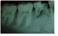

We have two sets of teeth. Primary dentition consists of 20 teeth and secondary or permanent dentition 32 teeth. The roots of primary teeth will usually dissolve or resorb between the age of 6 to 14, to allow these teeth to drop off and new teeth to come up.


I have encountered cases where the patients have congenitally missing teeth. The medical term for this condition is called 'hypodontia'. The common teeth that are missing in the permanent dentition are upper lateral incisors and lower second premolars. Sometimes their primary/deciduous teeth still remain in the jaws. The patient usually is not aware that the tooth has not been replaced by a permanent one, unless being told by his or her dentist during a routine check-up. I usually confirmed this occurence by taking a radiograph.

There are also cases where the patients complain of toothache due to large decay on these teeth. Sometimes, I perform a root canal treament, provided the root are not resorbed, in order to keep the tooth functioning in the oral cavity for as long as possible, especially for a young patient. However, the prognosis maybe be guarded. It is also possible that the retained primary tooth is fused to the jaw bone (ankylosed). I supposed one will just have to accept the fact that he or she is born with less teeth and ultimately, will need dental implants.


Friday, March 26, 2010

My Friday morning routine


I have been teaching dental students at the Dental Faculty, Universiti Kebangsaan Malaysia, for the past eight years. This is the place where students are trained to become one of us, the dentists.


I supervise them in the Restorative clinic. This clinic is in a large room with about twenty cubicles. Each cubicle has a dental chair. Dental treatment here is free and is open to public. A registered patient will be treated by dental students, who usually work in pairs. Most of the Year Four students are good and eager to learn. They remind me of my dental student's days. This is the time when you feel like a 'sponge' and you are required to absorb as much knowledge as your brain can take. Glad that this chapter of my life have ended. My duty is to guide them and check their work, step by step. I always tell my students that it is better to make a mistake here in dental school than when they are out on their own because they can learn from their mistake and I will be there to help and rectify it. That's the purpose of supervision.


Eight years is a long time. Perhaps, I should tell the Deputy Dean, who is a good friend of mine that it is time for me to close this chapter of my life too....

Thursday, March 25, 2010

X-ray Vision


As a dentist, I sometimes wish I have X-ray vision so that I can see through those teeth and jaws...


Radiography is essential in dental practice. Radiographs are useful in detecting tooth decay during a routine dental check-up, especially decay under an old filling (secondary caries) and decay in between teeth (interproximal caries). It is better to detect interproximal cavities early to avoid a more complex and expensive dental treatment when decay becomes large. Some of these cavities are consealed and difficult to detect by just doing an intraoral examination. After explaining this to my patients, they usually agree to do the radiographic examination. Most dental procedures require taking radiographs. For each root canal treatment, at least three radiographs are required.


Many are concerned about ionizing radiation from the dental X-ray. X-ray machines nowadays are more sophisticated. I assure my patients that the radiation dosage is very low, which is 1-8 microSv for an intraoral radiograph (Bitewing/Periapical). We are exposed to more radiation from natural sources everyday, for example, cosmic ray, gamma radiation, foodstuff etc. To put you in perspective, the average annual dose of radiation from cosmic rays and gamma radiation are 300microSv and 400microSv, respectively.


X-ray facilites in private dental clinics are regulated by The Ministry of Health , with specific guidelines, for example, minimum size of the X-ray or treatment room and lead-lined walls of the room, to ensure safety radiation.


Wednesday, March 24, 2010

Give Root Canal Treatment a Second Chance.

It is not uncommon for a patient to refuse root canal treatment (RCT) when he or she needs one because of a past RCT that has failed and ended up losing the tooth. I am always disappointed to hear this.

I was taught in my Masters programme that RCT has 70-90% of favourable treatment outcome. Perhaps this patient is one of the few unlucky ones. I will not bore you with the long list of reasons that influence the outcome of RCT, but I just want to emphasize here that if the treatment is carried out under acceptable standard of care, it will usually solve the patient's painful experience due to the dental infection. If the tooth is still restorable, RCT is definitely the better option than removing it. Losing your teeth will also lead to a set of other dental problems later on.

So, to these people who refuse RCT, don't let that just one bad experience influences your decision, give RCT a second chance!

It is also possible to retreat or repeat the RCT on a tooth when the first RCT does not have a favourable outcome.