Wednesday, March 2, 2011

Perforation Repair


When I was at Penn someone said that you have not been doing enough root canals if you haven't perforated a tooth. I am not sure if that is an accurate statement. At least not when you are working with a microscope.

This is a case of a lower right first molar that was left open for drainage by a previous dentist and a perforation occurred at the furcation during cavity access. The patient failed to return for further treatment and the tooth was left opened for more than two years. Hence, the large radiolucency on both roots and furcal area. A discharging sinus was present at lingual sulcus. I dont agree with the practice of leaving the tooth open for drainage and sending the patient home because it will cause more damage than good with all the contamination.

RCT was initiated and the furcal perforation was repaired with MTA. CaOH was left in the canals for three months. When there
was evidence of healing/ bony repair, canals were obturated. The six month review radiograph showed complete periapical healing and the radiolucency around the furcal area reduced in size. Although the lingual sinus has completely closed, I expect to see more healing at the furcation when I review this case in six months.

Saturday, February 26, 2011

Willkommen in Malaysia!


Chris is a friend from Germany, who came to visit Malaysia this week. We have a common friend and this is his first trip to Kuala Lumpur. He is a very talented person because not only he practises as a dentist, he is also an architect, a yoga teacher teacher and a professional photographer (This is his website: www.dade.de )

So, my nurses and I were very excited when he expressed his interest to visit my clinic in Petaling Jaya because he wanted to see how I worked with the operating microscope on root canal treatment. I scheduled a RCT on a lower first molar. My patient didn't mind having an observer during his treatment. He asked us if there was any dental equipment in my dental office that has German technology. The first thing that came to my mind was my BMW, which I simply loved, but my car was not a dental equipment. Looked like Dr Chris was
the only one there that was made in Germany.

At first, I was a little bit nervous. The last time another dentist looked over my shoulder and watched me worked was when I was in dental school. The treatment went smoothly and I was satisfied with my work, as shown in the final radiograph, which I always regarded it as my "report card".

The next patient came in for a routine check-up. She was such a good sport. Chris and I had a good discussion on the best dental treatment plan for her and she even helped us snapped some photos after her treatment.

I hope my friend enjoyed the rest of his stay in Malaysia and that he will have many good reasons to visit Kuala Lumpur again.

Thursday, February 17, 2011

Kong Hei Fatt Choy!


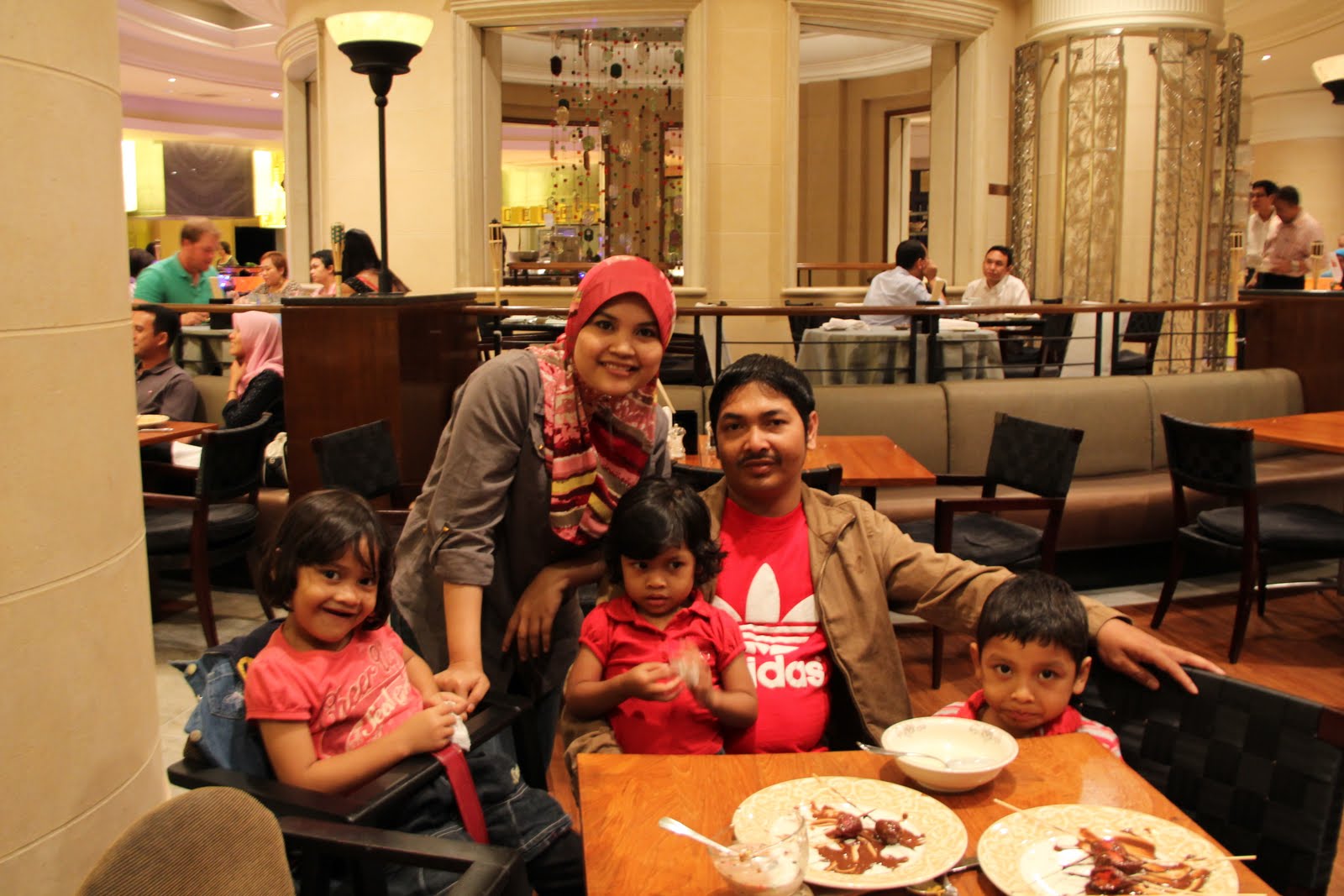
Today is the fifteenth day or the last day of Chinese New Year, also known as Chap Goh Meh. It was a great new year celebration, and I think the highlight of this CNY was our Yee Sang dinner at the Toh Yuen Restaurant, PJ Hilton. I kept my promise to treat my nurses, Asma and Banun, to an eight course dinner. It was a joyous occasion, attended by Banun and Asma, accompanied by their respective families, my friends Dalia, Matt, Shiamala and Azura. That made up to a table of nine adults and four children. The staff of the restaurant was very nice to offer us a room with beautiful oriental deco. We had fun toasting yee sang as Matt explained the significance of this Chinese New Year tradition. We ordered the Happiness Set Dinner. The food was halal of course and very tasty. I can tell the chef had put in a lot of effort preparing all the special dishes and I love the roasted chicken most. It was a nice surprise for me to see the kids, aged between three to nine year old, enjoying every course! The servings were large and it was just enough for all of us. My only complaint was the waitresses there were not very attentive and friendly. Maybe, they were too busy serving the Datuks and VIPs in the restaurant that night.

I take this opportunity to wish all friends of Soo Dental Surgery a happy and prosperous Chinese New Year and may the bunny brings lots of joy, good fortune and good health throughout the whole year. Kong Hei Fatt Choy!

Sunday, January 2, 2011

Analyzing toothache


A toothache that brings a patient to the dental office may either be inflammation of the dental pulp or when the pulp is already dead (necrotic). Although lack of pain does not represent a healthy pulp. Pulpal pain symptoms occur with different intensities, e.g. mild, moderate, or severe which is governed by frequency of firing, number of nerves and type of nerve fibres.

A-delta fibers are fast-conducting and evoke a rapid, sharp, lancinating pain reaction, and C-fibers are slow-conducting and cause a slow, dull, crawling pain. C-fibers have a higher excitability threshold than the A-delta fibers. A-delta fibers are located the periphery of the pulp, mostly at the coronal portion, and C-fibers are found centrally in the pulp proper.

Severity and duration of pain is related to the status of pulp pathosis.

Pulp inflammation may be reversible (reversible pulpitis) or irreversible (irreversible pulpitis). Information provided by the patient on nature of pain is important to differentiate between reversible pulpitis and irreversible pulpitis. Standard dental procedures on a tooth with reversible pulpitis is expected to reverse the inflammation of the pulp to a healthy state, whereas tooth with irreversible pulpitis will require an aseptic removal of the entire pulp (root canal treatment).

Symptoms associated with reversible pulpitis: No pain or sharp pain (mild) on contact with cold/hot liquid or sugary substances. Pain goes away when stimulant is removed. Peripheral A-delta fibers are stimulated.

Symptoms associated with irreversible pulpitis: No pain or pain on contact with cold and especially hot liquid, relieved by cold liquid. Pain is severe, throbbing, spontaneous, lingering and wakes the pain from sleep at night. There is usually a past history of pain and severe pain that is difficult to locate accurately (referred pain). The centrally located C-fibers are involved with these pain symptoms.

At this stage, the dentist should have an idea of the diagnosis of the tooth in question. The subsequent step is to determine the tooth that caused pain. A series of test that follows (cold test, heat test and electric pulp test) will determine whether the tooth is alive (vital) or dead (necrotic). These test are rather subjective and are not sensitive to differentiate between reversible pulpitis and irreversible pulpitis. One study shows that 83% of the teeth with a necrotic pulp were identified as necrotic by the cold test, while 93% of the teeth with vital pulp were identified as vital by the cold test. 86% of the teeth with necrotic pulp were identified as non vital by the heat test, while only 41% of the vital teeth were identified as vital by the heat test. For the Electric Pulp Test 72% of the teeth with necrotic pulp were identified as non vital, while 93% vital pulp were identified as vital in EPT test.

Percussion (tapping on tooth with a metal instrument) and palpation, are to evaluate periodontal inflammation. Necrotic or irreversibly inflamed pulp can lead to inflammation of the periodontal ligament. Inflammation of a tooth with referred pain remains confined within the pulp. The inflammatory process has not yet extended into the periapical region to engage the tactile fibers to give the clinical percussion sign. Sites of referred pain are always the posterior teeth and always unilateral, involving only one tooth in either maxilla or mandible.

In some situations, even the most experienced dentist may not be able to detect the culprit tooth or arriving to a diagnosis. The rule of thumb is to adapt the "wait and see" policy and in most cases, signs and symptoms will be more clearer in the subsequent appointments.


Wednesday, December 8, 2010

The missed canal: hidden or neglected?


The pre-op pa showed a large pa lesion associated with 25. The less centered canal at the apical third indicating the presence of another canal. That is where all the bacteria are harboring. Since there is no post or crown on this tooth, retreating this tooth may not be a difficult task.

How would a periapical lesion on this upper left second premolar heal? If one can get to the apical third to remove the microbes. Upon removal of gutta percha, buccal canal was instrumented to working length, which is 19mm. Palatal canal was detected. It was a (1-2) canal configuration, which the bifurcation occuring at 15mm.

I have cleaned and shaped the palatal canal, placed in CaOH and wait for healing to take place.

Monday, November 1, 2010

Peri-apical healing of a cracked tooth


Tooth 25 had a vertical fracture line that extended from mesial marginal ridge and all the way down into the orifice of palatal canal. I thought this was a hopeless case and the patient was very keen to keep the tooth. After dressing with CaOH for two weeks, there was still exudation in palatal canal. Obturation was done after six weeks of CaOH and when both canals were dry. A six-month review showed evidence of peri-apical healing.